
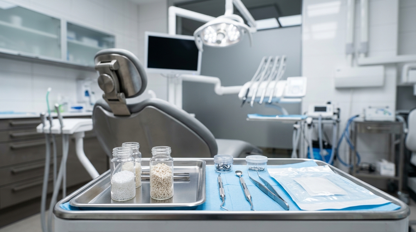
Every implant case is ultimately a bone management case. Whether you’re performing socket preservation after an atraumatic extraction or staging a major horizontal augmentation before placing a fixture, the graft material you select shapes the biology of the healing site, the resorption timeline, and—critically—when you can confidently proceed to implant placement. Understanding the four principal graft classes allows you to tailor your material choice to the clinical scenario rather than defaulting to habit.
The Four Classes at a Glance
Bone graft materials are categorized by their origin and mechanism of action. Each class offers a distinct biological profile that determines how the host site responds and how quickly it matures to implant-ready density.
- Autograft — harvested from the patient’s own body; osteogenic, osteoinductive, and osteoconductive.
- Allograft — derived from human donors; primarily osteoinductive and osteoconductive depending on processing.
- Xenograft — of animal origin, most commonly bovine; osteoconductive scaffold with slow, predictable resorption.
- Alloplast — fully synthetic; osteoconductive, with variable resorption determined by composition.
No single class dominates every situation. The ideal choice integrates defect morphology, patient biology, timeline constraints, and your confidence with the handling characteristics of the material.
Autograft: The Biological Benchmark
Autogenous bone remains the reference against which all other materials are judged, because it is the only class that delivers all three osteogenic mechanisms in a single graft. Harvested cortical or cancellous bone contains viable osteoblasts and osteoprogenitor cells (osteogenesis), growth factors including bone morphogenetic proteins (osteoinduction), and a collagen-mineral scaffold for cellular ingrowth (osteoconduction).
In implant dentistry, intraoral donor sites—the retromolar pad, the ascending ramus, the chin symphysis, or the implant osteotomy itself in the form of bone chips—provide quantities sufficient for localized socket preservation and small horizontal defects. For larger augmentations, calvarial or iliac crest harvest may be necessary, substantially increasing procedural complexity and patient morbidity.
The practical trade-off is resorption. Cortical block autografts are predictable for contour augmentation but experience variable surface resorption during the early healing phase, and particulate autograft resorbs comparatively quickly—within weeks to a few months if used alone. This rapid biology is an asset when speed matters, but it becomes a liability in defects requiring long-term volume maintenance. Many clinicians therefore combine autograft particulate with a slower-resorbing scaffold material to capture the osteoinductive advantage while preserving ridge volume through the maturation phase.
Handling-wise, autograft requires careful attention. Keep harvested material moist and viable: continuous saline irrigation during collection, storage in autologous blood or saline, and prompt placement are non-negotiable steps that directly affect cellular survival.
Allograft: Versatility and Broad Applicability
Processed cadaveric bone has become the most commonly used graft material in implant-focused oral surgery in North America, largely because it combines meaningful osteoinductive potential with broad availability and no donor-site morbidity. Two primary forms dominate clinical use:
- Demineralized freeze-dried bone allograft (DFDBA) exposes the collagen matrix and bound growth factors by removing the mineral phase, enhancing osteoinductive signaling. Its relatively rapid resorption—typically several months—makes it well-suited for socket preservation and contained defects where early bone formation is the priority.
- Freeze-dried bone allograft (FDBA) retains the mineral phase, providing a more durable osteoconductive scaffold with slower resorption. It is preferred when volume maintenance over a longer healing period matters more than early remodeling activity.
Rigorous donor screening and processing—including freeze-drying, irradiation, or chemical treatment—reduces immunogenic potential and renders allografts microbiologically safe. Disease transmission risk, while theoretically non-zero, is considered exceedingly low with tissue-bank-certified material processed under AATB standards.
From a handling standpoint, allograft particulate is forgiving. It hydrates readily with blood or saline, packs into sockets without excessive manipulation, and is available in a range of particle sizes. Coarser particles in the 750–1000 µm range are preferred in many socket preservation protocols to create interparticulate space for vascular ingrowth without early collapse of the graft mass.
Implant timing after allograft-based socket preservation generally falls in the 3–6 month range depending on defect size, patient healing capacity, and the blend of DFDBA versus FDBA used. Larger staged augmentations may require 6–9 months before fixture placement is appropriate.
Xenograft: The Slow-Resorbing Workhorse
Bovine-derived xenograft—most commonly deproteinized anorganic bovine bone mineral (DBBM)—has earned an enduring role in ridge preservation due to one defining characteristic: it resorbs very slowly, maintaining ridge volume for extended periods. The deproteinization process removes the organic component while preserving the mineral architecture, which closely mirrors the porosity and interconnectivity of human cancellous bone.
Because it lacks viable cells and growth factors, xenograft is purely osteoconductive—it provides a passive scaffold colonized gradually by host vasculature and osteoblasts. Resorption timelines are significantly longer than autograft or DFDBA; residual xenograft particles are routinely identified histologically in core biopsies taken years after placement. This slow resorption profile is a genuine asset for ridge preservation cases where long-term dimensional stability outweighs any interest in rapid remodeling.
The implant scheduling implication is important: longer healing windows—typically 6 months or more after socket preservation—are generally needed before bone density at the fixture site is optimal. Residual mineral particles incorporated within a maturing trabecular matrix do not impede osseointegration when present within the osteotomy path, but many clinicians prefer to confine xenograft to the peripheral areas of a socket, leaving the central core for faster-maturing bone.
Xenograft handling is straightforward. Products are available as dry granules or pre-hydrated collagen composites. Collagen-bound formats offer improved initial stability in fresh extraction sockets and require less condensation to maintain socket fill. Porcine-derived xenografts have also gained clinical traction, offering a somewhat faster resorption profile than bovine DBBM while retaining comparable scaffold architecture.
Alloplast: Synthetic Options and Their Role
Alloplastic materials are entirely synthetic, eliminating concerns about donor variability, disease transmission, and religious or cultural objections to biologically derived products. Several distinct compositions are in active clinical use:
- Beta-tricalcium phosphate (β-TCP) resorbs relatively quickly—faster than hydroxyapatite—and is often selected for sites where complete resorption before implant placement is preferred and a defined short healing window is planned.
- Hydroxyapatite (HA), whether sintered or non-sintered, provides a stable osteoconductive scaffold. Sintered HA is highly crystalline and resorbs very slowly, comparable to xenograft in long-term persistence.
- Biphasic calcium phosphate (BCP) combines β-TCP and HA in varying ratios, allowing modulation of resorption rate based on the HA:β-TCP ratio selected for the specific clinical objective.
- Bioactive glass bonds directly to bone through a surface hydroxycarbonate apatite layer and has demonstrated favorable results in certain localized alveolar defect applications.
Alloplasts perform best in well-contained defects with adequate bony walls to support the scaffold. They tend to be less forgiving in large, open augmentations without additional membrane support or co-grafting with biologically active material. Because they lack intrinsic osteoinductive capacity, their clinical performance depends entirely on adequate host cell recruitment into the scaffold.
Resorption Timelines and Implant Scheduling
When planning implant timing after augmentation, think in terms of material class, not a universal waiting period:
- Autograft alone: rapid remodeling; implant placement often appropriate at 3–4 months for socket preservation, 4–6 months for integrated block grafts.
- DFDBA-dominant mixes: a 3–5 month target range is typical for socket preservation in healthy patients.
- FDBA or FDBA/DFDBA blends: 4–6 months for preserved sockets; extended for staged augmentations.
- Xenograft (DBBM): 6 months or beyond for standard socket preservation; longer for larger defect volumes.
- Alloplast (β-TCP): 3–5 months; HA-dominant or high-HA BCP products follow xenograft-range timelines.
These windows assume a healthy non-smoking patient with no significant systemic comorbidities. Uncontrolled diabetes, active smoking, antiresorptive medications, and prior site radiation compress or extend these estimates meaningfully and should factor explicitly into your preoperative conversation.
Matching Material to Scenario
For routine socket preservation following atraumatic single-tooth extraction, an FDBA particulate under a resorbable collagen membrane represents a well-supported, broadly available protocol with predictable outcomes. When an osteoinductive stimulus is clinically beneficial—a compromised socket, a thin buccal plate, a patient with moderate healing demands—incorporating autograft chips or DFDBA adds biological potency without sacrificing handling ease.
For staged horizontal ridge augmentation requiring significant volume gain, cortical block autograft or a cortical allograft block with particulate fill under a resorbable or non-resorbable membrane provides a reliable architecture. Adding xenograft to the peripheral layer can slow surface resorption during the maturation window and preserve contour while the inner core consolidates.
For patients with cultural or religious objections to animal-derived products, alloplastic or allograft-only protocols deliver equivalent biological performance in most socket preservation applications without compromise on predictability.
The graft material sets the stage—but implant success depends on what you place into a well-prepared site, how the membrane protects the space, and how precisely the fixture engages mature, adequately dense bone. Matching material class to clinical scenario rather than defaulting to a single product for every case is what separates a thoughtful augmentation strategy from a routine one.